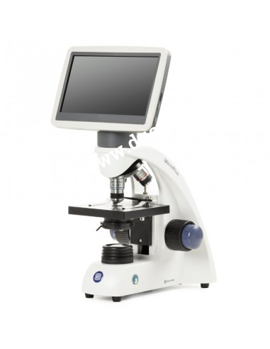
Exploring Electron Microscopes: Superior Tools in Scientific Research
I. Introduction to Electron Microscopes
Electron microscopes are crucial tools in modern scientific research. With the ability to magnify up to millions of times, electron microscopes allow scientists to explore the microstructures of materials and organisms, opening new doors in various research fields. This article will help you understand electron microscopes, their structure, principles of operation, applications, and their outstanding advantages.

II. Structure and Operation of Electron Microscopes
Detailed Structure
Electron microscopes (EM) are vital in scientific and medical research, enabling high magnification and resolution observation of microstructures. There are two main types of electron microscopes: Scanning Electron Microscope (SEM) and Transmission Electron Microscope (TEM).
Structure of TEM:
- Electron Gun: Generates an electron beam.
- Lens System: Includes condenser lenses, objective lenses, intermediate lenses, and projector lenses.
- Sample Chamber: Where the sample is placed for observation.
- Fluorescent Screen: Displays the image of the sample.
Structure of SEM:
- Electron Gun: Generates an electron beam.
- Condenser and Objective Lenses: Focus and adjust the electron beam.
- Sample Chamber: Where the sample is placed for scanning.
- Detector: Collects emitted electrons from the sample and converts them into images on the screen.
Electron microscopes have a complex structure with essential components such as the electron gun, lens systems, and detectors, which contribute to producing detailed images of microstructures not visible to the naked eye. These components are crucial for high magnification and resolution, serving scientific research and medical applications.
III. Operating Principles
Electron microscopes operate based on the principle of using an electron beam instead of light to create images. The electron beam is emitted from an electron source, then directed and focused by the electron lens system. When the electron beam interacts with the sample, the reflected or transmitted signals are collected and converted into images on a display screen.
Types of Electron Microscopes
-
Scanning Electron Microscope (SEM)
- SEM uses an electron beam to scan the surface of the sample, creating high-resolution three-dimensional images. SEM is suitable for observing the surface and external structures of samples.
-
Transmission Electron Microscope (TEM)
- TEM uses an electron beam transmitted through the sample to create detailed images of internal structures. TEM offers higher resolution than SEM and is commonly used for studying the internal microstructures of samples.
IV. Applications of Electron Microscopes
Electron microscopes have significant applications in various scientific and technological fields:
In Biological Research
- Electron microscopes help biologists observe cell structures, viruses, and other microorganisms at the atomic level, providing deeper insights into biological processes.
In Materials Science
- Electron microscopes enable the analysis of microstructures of materials, improving their properties and performance. For example, studying the structure of semiconductor materials can lead to the development of higher-performance electronic devices.
In Medical Applications
- Electron microscopes are used to observe tissues and diseased cells, aiding in the diagnosis and treatment of various medical conditions. Scientists can examine detailed structures of cancerous tissues to develop new treatment methods.
V. Advantages and Limitations of Electron Microscopes
Outstanding Advantages
- High Resolution: Electron microscopes can magnify up to millions of times, allowing detailed observation of ultra-small structures.
- Diverse Applications: Used in many scientific and technological fields.
- Detailed Imaging: Provides sharp and detailed images.
Limitations and Challenges
- High Cost: Electron microscopes are expensive.
- Technical Expertise Required: Users need specialized knowledge and skills.
- Special Operating Conditions: Requires a dust-free and low-vibration environment.
VI. How to Use Electron Microscopes
Basic Guidelines
- Sample Preparation: Carefully prepare samples to ensure image quality.
- Microscope Setup: Adjust the microscope settings to match the sample.
- Observation and Imaging: Use the microscope to observe and capture images of the details to be studied.
Usage Notes
- Sample Preservation: Ensure samples are properly preserved to prevent damage.
- Microscope Maintenance: Regularly clean the microscope components to ensure optimal performance.
Prominent Electron Microscope Brands
- Euromex – Netherlands: Offers a range of high-quality microscopes.
- Zeiss: A leading brand in electron microscopy.
- Hitachi: Known for high-quality electron microscopes.
- Thermo Fisher Scientific: Provides various advanced electron microscopes.
- JEOL: A reputable brand with a wide range of electron microscopes.
- FEI Company: Known for modern electron microscopy solutions.
VII. Conclusion
Electron microscopes are indispensable tools in modern scientific research, enabling scientists to explore the wonders of the micro-level. With high resolution and diverse applications, electron microscopes significantly contribute to the advancement of many scientific and technological fields.
VIII. Frequently Asked Questions
-
How do electron microscopes work?
- Electron microscopes use an electron beam to create images of the sample. The electron beam interacts with the sample, producing signals that are then converted into images on a display screen.
-
What are the advantages of electron microscopes over optical microscopes?
- Electron microscopes offer much higher resolution compared to optical microscopes, allowing for detailed observation of ultra-small structures that optical microscopes cannot see.
-
How much does an electron microscope cost?
- The cost of an electron microscope can range from tens of thousands to millions of USD, depending on the type and features of the microscope.
-
What are common applications of electron microscopes?
- Electron microscopes are widely used in biological research, materials science, medicine, and many other scientific fields.
IX. Where to Buy Electron Microscopes
Duc Duong Scientific and Technical Company
With many years of experience, Duc Duong has increasingly established its brand and reputation in providing laboratory furniture and equipment, having executed numerous projects for laboratories, microbiology research rooms, molecular biology, tissue culture, biosafety, environmental, food, pharmaceutical, and aquaculture sectors.
Contact us today for consultation: [Contact Information]
Or reach us at the hotline below:
ĐỨC DƯƠNG TECHNOLOGY SCIENCE COMPANY LTD
Address: 1014/67 Tân Kỳ Tân Quý, Bình Hưng Hòa, Bình Tân, HCM
Tel: (028) 3762 8042 - 3762 8043 - 3750 8514 - 3750 8793
Fax: 028 37628043
Email: ducduong@ducduongco.com
Website: www.ducduongco.com
Comments
Relative post
- Different for Food autoclave and Medical autoclave
- Autoclave Sterilizers: A Comprehensive Guide to Construction and Operation
- DUC DUONG TECHNOLOGY SCIENCE CO., LTD Participates in the 17th Medical and Pharmaceutical Exhibition in HCMC
- Necessary Experience When Using Microscopes
- Duc Duong Company will participate in Analytica Vietnam Exhibition in April 2019
- AUTOCLAVE STERILIZER - SHINJINENG


